
What a Skeletal Scan Can and Cannot Diagnose
If someone tells you to “get a skeletal scan,” the first question should be: which kind?
The phrase sounds specific, but in real medical use it can refer to several different imaging tests. A nuclear medicine bone scan, a DEXA scan, a skeletal survey, CT, MRI and standard X-rays can all look at the skeleton in different ways. They do not answer the same question.
That distinction matters because a skeletal scan might help detect abnormal bone activity, measure bone mineral density, locate a fracture or assess body composition, depending on the test. It may also be the wrong tool if you need to diagnose a soft tissue injury, identify the exact cause of pain or confirm a specific cancer type.
Below is a practical guide to what a skeletal scan can and cannot diagnose, plus how to choose the right test for the question you are trying to answer.
What does “skeletal scan” usually mean?
“Skeletal scan” is not one standardized test name. Patients often use it as a general phrase for imaging that evaluates bones. Clinicians usually use more precise terms.
The most common possibilities are:
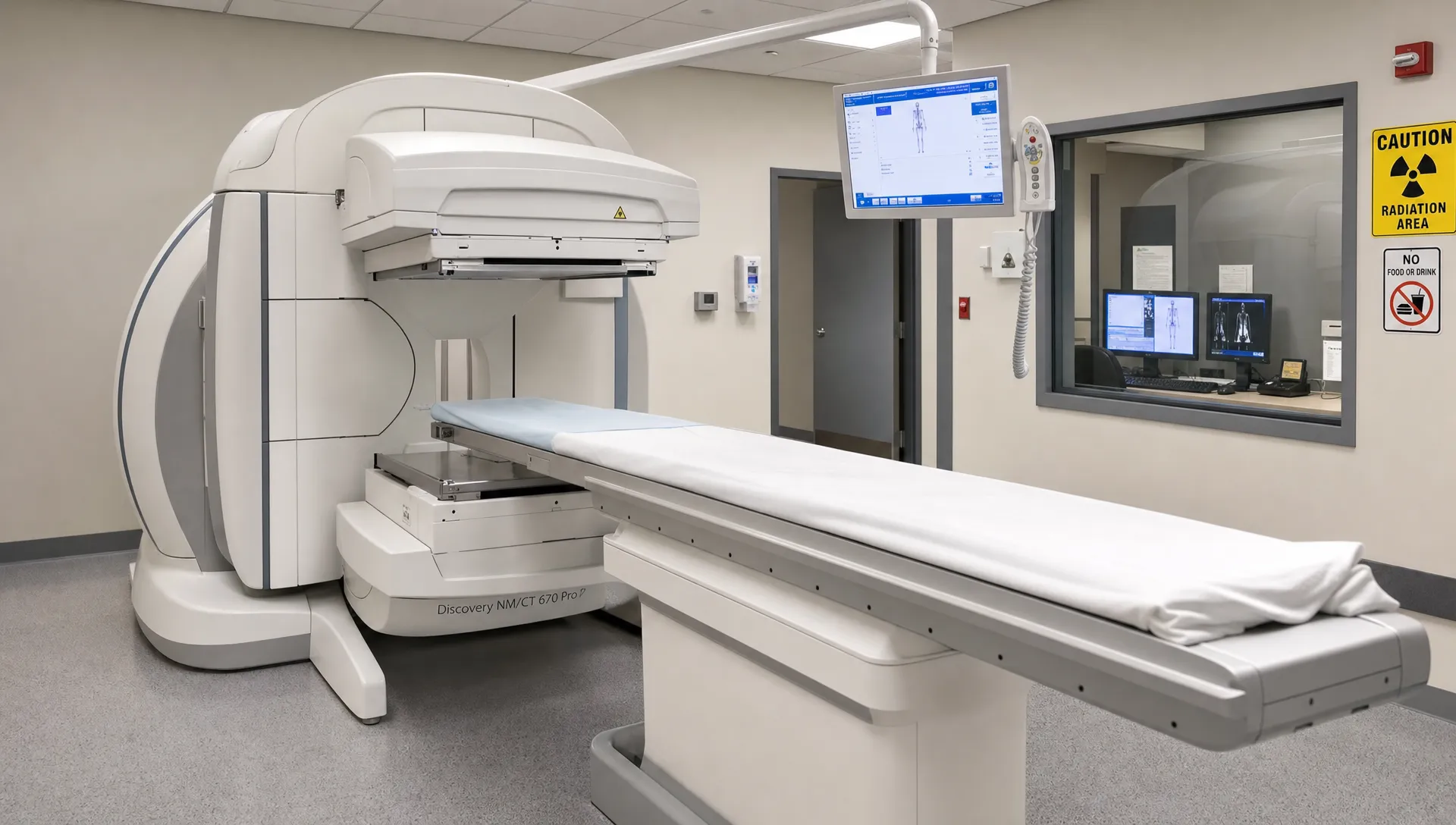
- Nuclear medicine bone scan: Also called bone scintigraphy. A small amount of radioactive tracer is injected into a vein, then a camera detects areas of increased or decreased bone turnover.
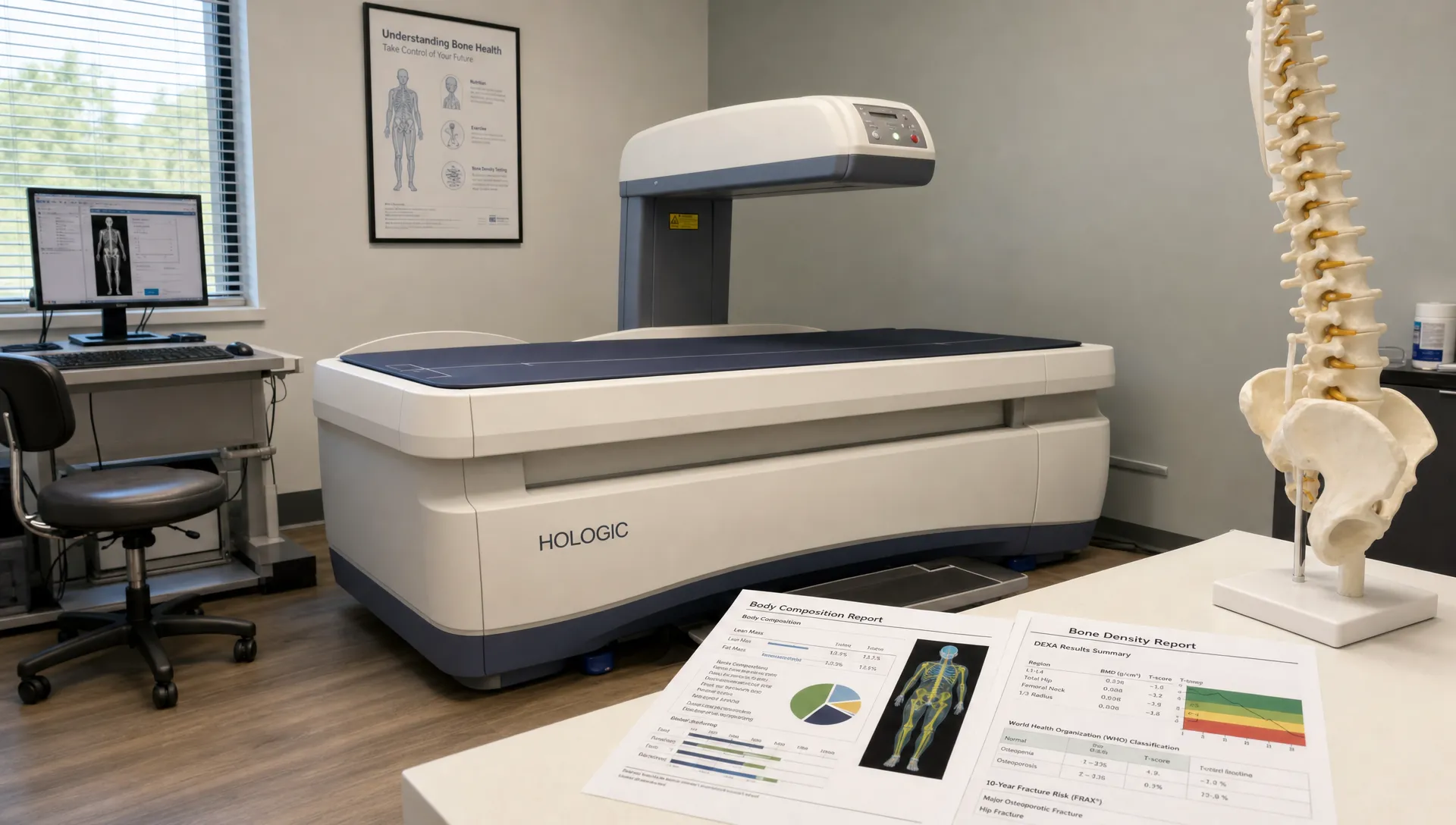
- DEXA scan: Also called DXA. A low-dose X-ray test that measures bone mineral density and, when performed as a body composition scan, fat mass, lean mass and other metrics.
- Skeletal survey: A series of X-rays of many bones, often used in specific settings such as suspected multiple myeloma, certain pediatric concerns or skeletal dysplasias.
- CT or MRI of the skeleton: More detailed imaging used when a clinician needs a closer look at a specific bone, joint, marrow space or surrounding soft tissue.
- Standard X-ray: A targeted image of one area, commonly used to evaluate suspected fractures, arthritis, alignment or obvious bone lesions.
If your goal is to understand bone density, osteoporosis risk or body composition, a DEXA scan is usually the relevant test. If your doctor is looking for abnormal bone activity throughout the body, such as possible metastatic disease or a hard-to-find stress fracture, they may mean a nuclear medicine bone scan. For a deeper comparison, DEXA SF has a separate guide on bone scan vs DEXA that explains how these tests differ.
What a skeletal scan can diagnose or help detect
A skeletal scan can be useful, but the answer depends on which scan is being performed. Some tests diagnose directly. Others identify suspicious findings that need confirmation.
Osteoporosis and low bone density
A DEXA scan is the standard imaging test for measuring bone mineral density. It can diagnose osteoporosis when the T-score meets established criteria, usually at the hip or lumbar spine, in the right clinical context. It can also identify low bone mass, often called osteopenia.
According to RadiologyInfo.org, DEXA is used to diagnose osteoporosis, assess fracture risk and monitor response to treatment. The U.S. Preventive Services Task Force recommends osteoporosis screening for women 65 and older, as well as postmenopausal women under 65 who are at increased risk, in its current osteoporosis screening recommendation.
A nuclear medicine bone scan is different. It may show areas of high bone turnover, but it does not measure bone mineral density and cannot diagnose osteoporosis in the way DEXA can.
Fractures and stress injuries
A skeletal scan can help identify fractures, but the best test depends on timing and location.
A standard X-ray is often the first test for a suspected fracture. It is fast and widely available, especially when there is a clear injury. Some stress fractures and small fractures may not appear on early X-rays.
A nuclear medicine bone scan can show increased bone activity from a stress fracture, sometimes before changes are obvious on X-ray. MRI is also commonly used for stress injuries because it can show bone marrow edema and nearby soft tissue damage. CT can be useful for fine bony detail, especially in complex areas.
A DEXA scan does not usually diagnose an acute fracture. Its role is different: it helps assess whether low bone density may be contributing to fracture risk.
Bone metastases and cancer-related bone changes
A nuclear medicine bone scan can help detect areas of abnormal bone turnover that may be caused by cancer spread to bone. This is one reason doctors order whole-body bone scans in people with certain cancers.
However, the scan does not diagnose cancer by itself. Increased tracer uptake can also happen with arthritis, healing fractures, infection or other bone conditions. A suspicious bone scan finding usually needs correlation with symptoms, lab tests, CT, MRI, PET imaging or biopsy.
A DEXA scan is not used to diagnose cancer or cancer spread. It can be useful for monitoring bone density in people whose cancer treatment affects bone health, but that is a different clinical question.
Arthritis and degenerative joint changes
X-rays can show structural signs of osteoarthritis, such as joint space narrowing, bone spurs and changes in alignment. CT may add detail in complex joints. MRI can show cartilage, menisci, ligaments, bone marrow changes and other soft tissues.
A nuclear medicine bone scan can show increased activity around arthritic joints, but that finding is not specific. It may help locate active areas when symptoms are unclear, but it usually cannot distinguish all possible causes on its own.
DEXA is not a diagnostic test for arthritis. It can sometimes be affected by degenerative changes in the spine, which is one reason interpretation matters.
Bone infection and inflammation
A nuclear medicine bone scan may help detect osteomyelitis, which is infection in bone, because infected areas can show increased tracer uptake. In many cases, MRI is preferred for local detail, especially when soft tissue involvement, abscess or marrow changes need to be evaluated.
A scan finding alone rarely completes the diagnosis. Clinicians may need blood tests, cultures, clinical examination and sometimes biopsy or aspiration.
Paget’s disease and abnormal bone remodeling
Paget’s disease of bone causes abnormal bone remodeling. A nuclear medicine bone scan can show the extent of active disease throughout the skeleton. X-rays and lab tests, including alkaline phosphatase in many cases, may also be used.
DEXA may be relevant if bone density needs to be assessed, but it is not the main test for mapping active Paget’s disease.
Muscle mass, fat mass and body composition
This is where DEXA has a separate advantage. A DEXA body composition scan can estimate total body fat percentage, lean mass, fat mass, visceral fat and left vs. right lean mass differences. Those data can be useful for athletes, people pursuing fat loss, people rebuilding muscle or anyone who wants more detail than scale weight can provide.
At DEXA SF, scans are used to measure body fat, muscle mass, bone density, visceral fat and asymmetries, with expert interpretation and guidance for fitness and nutrition goals. If you want a broader look at what body composition and bone metrics can show together, the guide on body and bone health from one scan is a helpful next step.
Quick comparison: which skeletal scan answers which question?
| Health question | Test commonly used | What it can show | Key limitation |
|---|---|---|---|
| Do I have low bone density or osteoporosis? | DEXA | Bone mineral density, T-score, Z-score | Does not diagnose cancer, infection or most acute fractures |
| Has cancer spread to bone? | Nuclear medicine bone scan, PET, CT or MRI | Suspicious areas of abnormal bone activity or lesions | Findings are not automatically cancer |
| Do I have a fracture? | X-ray, CT or MRI | Breaks, alignment, bone detail or marrow changes | Some early stress fractures may be missed on X-ray |
| Do I have a stress injury? | MRI, bone scan or X-ray | Bone stress reaction, fracture activity or structural change | Test choice depends on location and timing |
| What is my body fat and lean mass? | DEXA body composition scan | Fat mass, lean mass, body fat percentage, visceral fat | Does not diagnose disease from body composition alone |
| Do I have arthritis? | X-ray, MRI or CT | Joint structure, cartilage or bone changes | Imaging must match symptoms and exam findings |
What a skeletal scan cannot diagnose by itself
Imaging is powerful, but it is not a substitute for a complete diagnosis. A skeletal scan often provides evidence, not the entire answer.
It cannot always identify the exact cause of pain
Bone pain, joint pain and muscle pain can overlap. A scan may show arthritis, an old injury or an area of increased bone activity, but that does not always prove the finding is the source of symptoms.
For example, many adults have degenerative changes on imaging that are not the main cause of their pain. Clinicians interpret scan results alongside the physical exam, history, labs and response to treatment.
It cannot diagnose the type of cancer
A bone scan can detect suspicious bone activity, but it cannot tell you the exact cancer type. It also cannot reliably separate cancer from every benign cause of uptake.
A diagnosis of cancer usually requires a combination of imaging, medical history, laboratory tests and sometimes tissue sampling. If your doctor orders a skeletal scan for cancer evaluation, ask what result would lead to the next step.
It cannot replace lab tests for metabolic or hormonal problems
Low bone density may be related to age, menopause, medications, low body weight, nutrient deficiencies, thyroid or parathyroid problems, kidney disease or other factors. DEXA can measure bone density, but it cannot explain every cause.
If a scan shows low bone density, a clinician may order blood or urine tests to look for contributing conditions.
It cannot show everything about soft tissues
Tendons, ligaments, discs, muscles, cartilage and nerves are not always well evaluated by bone-focused imaging. MRI or ultrasound may be more appropriate when the suspected problem is soft tissue rather than bone.
This is especially relevant for sports injuries. A skeletal scan can help with bone stress questions, but it may miss the soft tissue reason an athlete has pain or impaired performance.
It cannot measure bone strength perfectly
DEXA measures bone mineral density, which is strongly related to fracture risk, but fracture risk is not determined by density alone. Age, prior fractures, medications, fall risk, family history, smoking, alcohol intake and certain medical conditions also matter.
That is why DEXA results are often interpreted with risk tools and clinical context. A person with osteopenia and a previous fragility fracture may have a different risk profile than someone with the same T-score and no other risk factors.
How DEXA results differ from other skeletal scan results
A DEXA report is more quantitative than many other skeletal imaging reports. Instead of simply describing what the radiologist sees, it gives measurements.
For bone health, a DEXA scan commonly includes bone mineral density values and scores. T-scores compare your bone density with that of a healthy young adult reference population. Z-scores compare your result with what is expected for someone of similar age and sex.
For body composition, a DEXA scan may include body fat percentage, total fat mass, lean mass, visceral fat estimate and regional comparisons. That makes it useful for tracking changes over time, especially when scale weight does not tell the full story.
The numbers still need context. Hydration, recent training, positioning and normal measurement variation can affect body composition tracking. Degenerative spine changes can also influence some bone density readings. If you already have results and want to understand the terminology, DEXA SF’s guide to DEXA scan interpretation explains common report metrics in plain language.
When should you ask about a skeletal scan?
You may want to discuss skeletal imaging with a clinician if you have unexplained bone pain, a suspected fracture, a history of cancer with new bone symptoms, risk factors for osteoporosis or a need to monitor bone health over time.
You may want a DEXA body composition scan if your main goal is to track fat loss, muscle gain, visceral fat or body symmetry more precisely than a scale, tape measure or body mass index can.
The right question helps determine the right test:
- If the question is “How dense are my bones?” ask about DEXA.
- If the question is “Is there abnormal bone activity across my skeleton?” your clinician may consider a nuclear medicine bone scan.
- If the question is “Did I break this bone?” X-ray, CT or MRI may be more appropriate.
- If the question is “What is my fat, muscle and visceral fat profile?” DEXA body composition is often the best fit.
How to prepare for a conversation with your clinician
Before asking for any skeletal scan, write down the problem you want the scan to answer. Include when symptoms started, what makes them better or worse, any injury history, cancer history, medications, fracture history and recent imaging.
If your goal is preventive bone health, ask whether your age, medical history, medications or family history make DEXA appropriate. If your goal is fitness or nutrition tracking, ask how often repeat scans make sense and which metrics matter most for your plan.
For people in San Francisco who want precise body composition and bone density data, DEXA SF provides professional DEXA scans with expert interpretation, fitness guidance and nutrition advice. That makes it a practical option when the question is about bone density, body fat, lean mass, visceral fat or asymmetry.
Frequently Asked Questions
Is a skeletal scan the same as a bone scan? Not always. Many people use “skeletal scan” to mean a nuclear medicine bone scan, but the phrase can also refer to DEXA, X-rays, CT, MRI or a skeletal survey. Ask which test is being ordered and what question it is meant to answer.
Can a skeletal scan diagnose osteoporosis? A DEXA scan can diagnose osteoporosis by measuring bone mineral density and calculating scores such as the T-score. A nuclear medicine bone scan does not diagnose osteoporosis because it does not measure bone density.
Can a skeletal scan detect cancer? A nuclear medicine bone scan can detect suspicious areas that may represent cancer spread to bone, but it cannot confirm the cancer type or prove that cancer is present by itself. Further testing may be needed.
Can a DEXA scan show fractures? DEXA is mainly used for bone density and body composition. It is not the usual test for diagnosing acute fractures. X-ray, CT or MRI is typically used when a fracture is suspected.
Is a skeletal scan painful? Most skeletal imaging tests are not painful, although some require lying still. A nuclear medicine bone scan involves an injection of tracer. A DEXA scan is quick, noninvasive and uses low-dose X-ray technology.
The bottom line
A skeletal scan can be very useful, but only when the test matches the question. DEXA can measure bone density and body composition with precision. A nuclear medicine bone scan can identify areas of abnormal bone activity. X-ray, CT and MRI each have their own strengths for fractures, joint disease, marrow changes and local anatomy.
If your goal is to understand bone density, body fat, muscle mass, visceral fat or body asymmetry, a DEXA scan is often the most relevant skeletal scan. If you are evaluating unexplained pain, possible cancer spread, infection or a fracture, your clinician may recommend a different imaging pathway.
To get precise DEXA measurements with expert interpretation in San Francisco, schedule a scan with DEXA SF and use your results to make more informed health, fitness and nutrition decisions.