
Bone Scan vs DEXA: What’s the Difference?
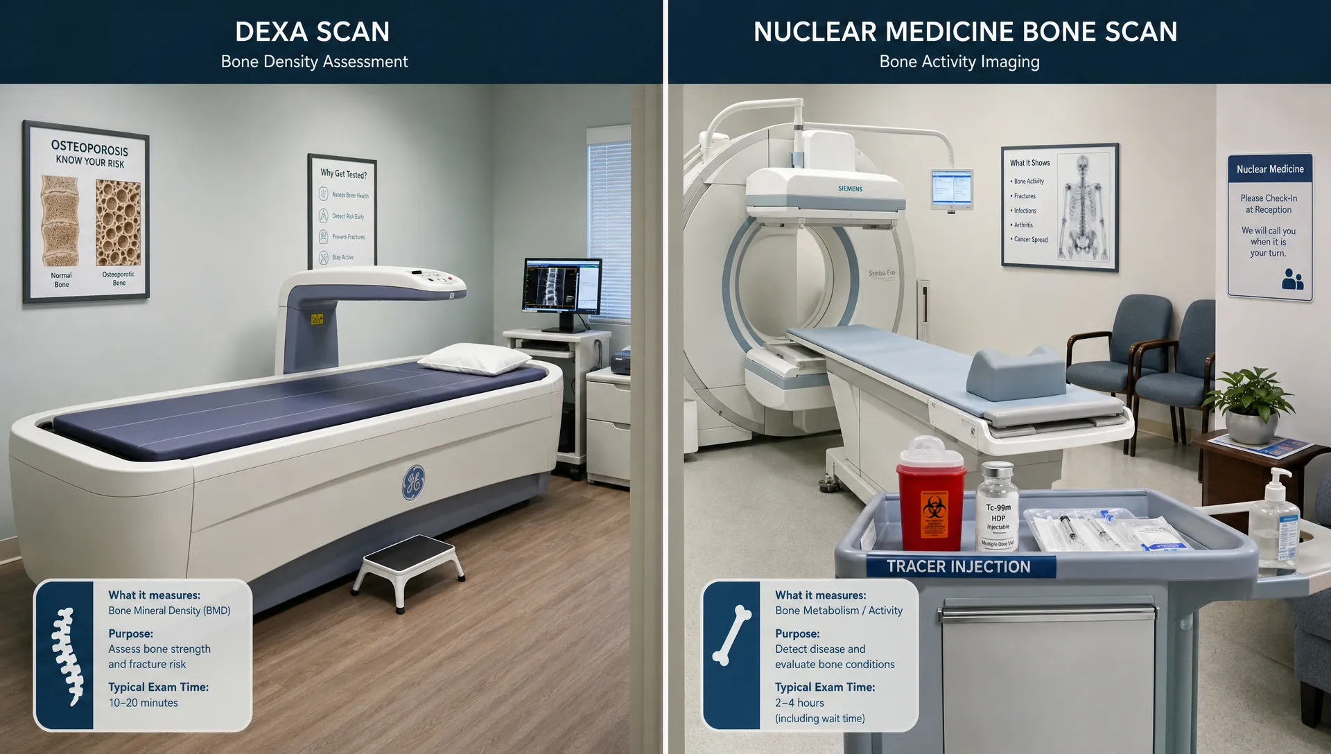
If you have been told to get a “bone scan,” it is worth pausing for one important reason: people use that phrase to mean different things. In everyday conversation, a bone scan might refer to any scan related to bones. In medical imaging, however, a bone scan usually means a nuclear medicine test that looks for abnormal activity in bone. A DEXA scan, also called DXA, is a different test that measures bone density and, in many cases, body composition.
That difference matters. A nuclear bone scan is often used to investigate a medical concern such as unexplained bone pain, a suspected injury, infection, or cancer spread to bone. A DEXA scan is used to measure bone mineral density, assess osteoporosis risk, and provide precise data on body fat, lean mass, visceral fat, and left vs. right muscle balance when performed as a body composition scan.
This guide breaks down bone scan vs DEXA in plain English, so you can understand what each test is for, what the results tell you, and which one is more relevant for your goals.
The short answer: bone scan and DEXA are not the same
A bone scan and a DEXA scan both involve imaging, but they answer very different questions.
A bone scan, in the strict medical sense, is a nuclear medicine exam. It uses a small amount of radioactive tracer that travels through the bloodstream and collects in areas where bone is actively repairing or changing. A camera then detects the tracer pattern. According to RadiologyInfo.org’s bone scan overview, this type of scan can help evaluate conditions such as fractures, infection, arthritis, and cancer that has spread to bone.
A DEXA scan uses low-dose X-rays to calculate bone mineral density. When performed for body composition, DEXA can also estimate total body fat percentage, lean mass, visceral fat, and regional differences between the left and right sides of the body. For a deeper look at bone density specifically, DEXA SF’s bone density scan guide explains what to expect and why tracking bone health matters.
Here is the simplest way to separate the two:
| If your main question is... | The more relevant test is usually... | Why |
|---|---|---|
| “How dense are my bones?” | DEXA | DEXA measures bone mineral density. |
| “Do I have osteoporosis or low bone mass?” | DEXA | DEXA is commonly used to assess osteoporosis risk. |
| “What is my body fat percentage and lean mass?” | DEXA | A body composition DEXA scan measures fat and lean tissue distribution. |
| “Is there abnormal bone activity from injury, infection, or disease?” | Bone scan | A nuclear bone scan highlights areas of increased or decreased bone activity. |
| “Is my right side stronger or more muscular than my left?” | DEXA | DEXA can show regional lean mass and asymmetries. |
Why the term “bone scan” causes confusion
The phrase “bone scan” sounds broad, so it is often used casually. Someone might say they are getting a bone scan when they mean a bone density test. A physician, radiologist, or nuclear medicine department may use “bone scan” to mean a specific nuclear medicine test.
If your paperwork says DEXA, DXA, bone densitometry, or bone density scan, it is referring to a DEXA scan. If it mentions nuclear medicine, radiotracer, Tc-99m, bone scintigraphy, or a camera scan after an injection, it is referring to a nuclear bone scan.
When in doubt, ask the ordering clinician or imaging center: “Is this a DEXA bone density scan or a nuclear medicine bone scan?” That one question can prevent a lot of confusion.
What is a nuclear medicine bone scan?
A nuclear medicine bone scan is designed to show bone metabolism, not bone density. In other words, it looks at where bone tissue is unusually active.
During the test, a tracer is injected into a vein. After a waiting period, the tracer collects in the skeleton. A special camera then captures images that show areas of higher or lower tracer uptake. These areas are sometimes called “hot spots” or “cold spots,” depending on the pattern.
A clinician may order a nuclear bone scan for reasons such as:
- Unexplained bone pain
- Suspected stress fracture or small fracture not clearly seen on X-ray
- Evaluation of cancer that may have spread to bone
- Possible bone infection
- Assessment of certain inflammatory or degenerative bone conditions
- Looking at multiple areas of the skeleton in one exam
A bone scan can be very useful, but it is not a general wellness or body composition test. It also does not directly tell you your bone mineral density, body fat percentage, visceral fat, or muscle mass.
What is a DEXA scan?
DEXA stands for dual-energy X-ray absorptiometry. As RadiologyInfo.org explains in its DEXA overview, DEXA uses two X-ray beams at different energy levels to estimate bone mineral density. It is widely used because it is quick, noninvasive, and involves very low radiation exposure compared with many other imaging tests.
In a clinical bone density context, DEXA often focuses on areas such as the hip and spine because those sites are important for osteoporosis and fracture risk assessment. In a body composition context, DEXA can scan the whole body and provide a detailed breakdown of fat mass, lean mass, bone mass, and regional distribution.
At DEXA SF, a DEXA scan can help measure:
- Body fat percentage
- Lean mass and muscle distribution
- Bone density
- Visceral fat
- Left vs. right asymmetries
- Metabolic data insights
The value is not just the scan itself. For people working on fitness, nutrition, longevity, or performance goals, DEXA results can turn vague progress into measurable data. Instead of relying only on scale weight, mirror checks, or wearable estimates, you can see what is actually changing in fat mass, lean mass, and bone metrics.
If you are comparing local options, this guide to what to expect from a DEXA scan in San Francisco covers practical details like use cases, timing, and how often people may choose to scan.
Bone scan vs DEXA: side-by-side comparison
The easiest way to understand the difference is to compare what each scan is built to do.
| Category | Nuclear medicine bone scan | DEXA scan |
|---|---|---|
| Main purpose | Detect abnormal bone activity | Measure bone density and body composition |
| What it shows | Areas of increased or decreased tracer uptake | Bone mineral density, fat mass, lean mass, and regional composition depending on protocol |
| Common use | Medical evaluation of pain, injury, infection, arthritis, or cancer spread | Osteoporosis risk assessment, body composition tracking, fitness and nutrition progress |
| Measures bone density? | No, not directly | Yes |
| Measures body fat? | No | Yes, with a body composition DEXA scan |
| Measures muscle mass? | No | Estimates lean mass by region and total body |
| Uses injection? | Yes, a radiotracer injection | No injection |
| Scan experience | Often involves injection, waiting time, then imaging | Typically quick and noninvasive |
| Result type | Images showing tracer uptake patterns | Numeric report with density and composition metrics |
| Best for | Medical investigation of bone activity | Quantifying bone health, fat, muscle, visceral fat, and asymmetry |
Which scan do you need?
The right test depends on the question you are trying to answer.
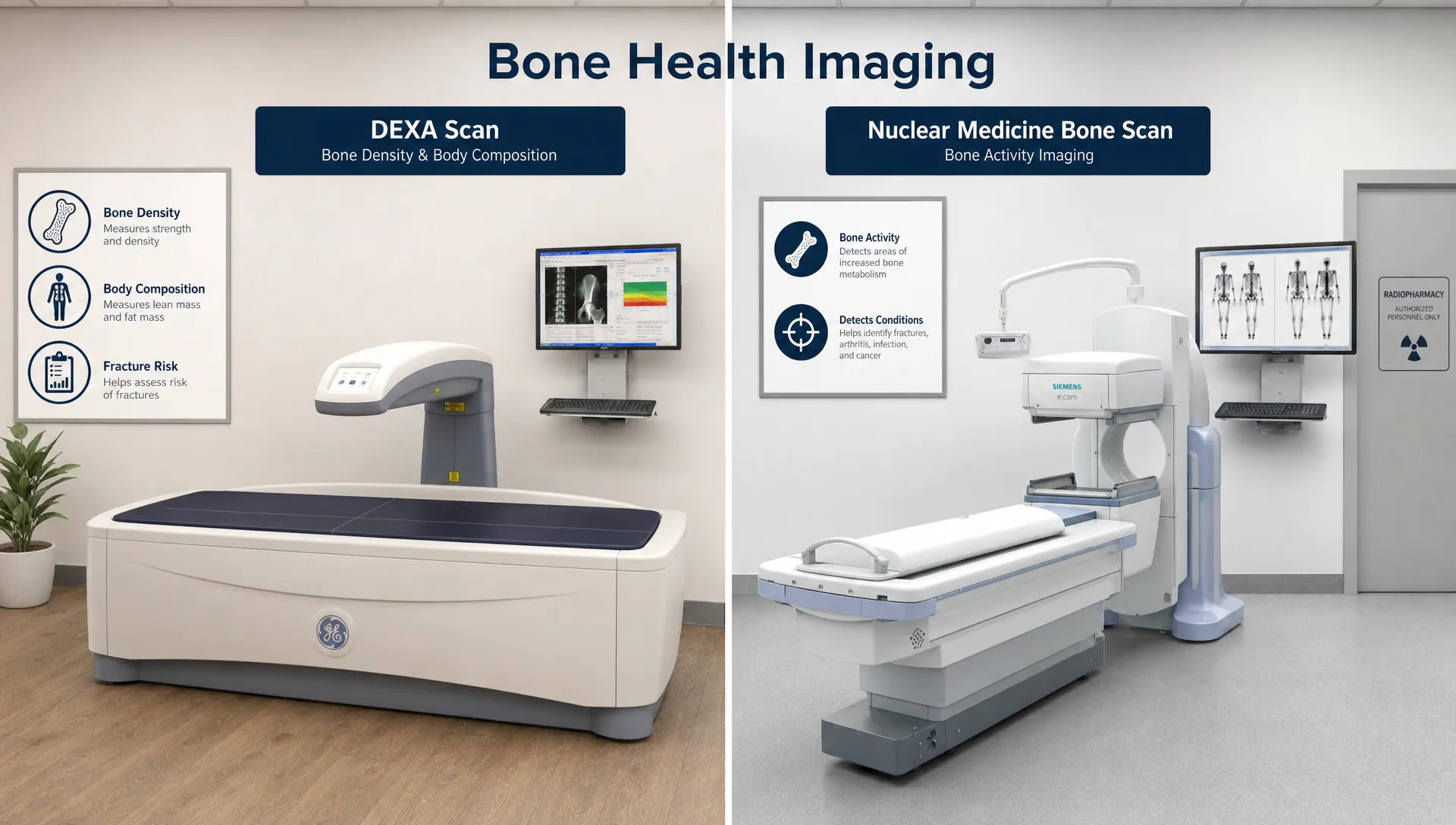
If your goal is to understand bone density, osteoporosis risk, or changes in bone mass over time, DEXA is usually the relevant scan. It gives measurable bone mineral density data that can be tracked and interpreted in context.
If your goal is to understand body composition, DEXA is also the better fit. A standard nuclear bone scan will not tell you how much fat or muscle you have, where your fat is distributed, how much visceral fat you carry, or whether one side of your body has more lean mass than the other.
If your goal is to investigate unexplained bone pain, suspected cancer spread, infection, or certain injuries, a nuclear medicine bone scan may be appropriate, but that decision belongs with your clinician. In some cases, your clinician may choose MRI, CT, X-ray, PET, or another test instead, depending on your symptoms and history.
A practical rule of thumb:
| Your goal | More likely match |
|---|---|
| Track fat loss without guessing | DEXA |
| Measure lean mass gain | DEXA |
| Assess visceral fat | DEXA |
| Screen or monitor bone density | DEXA |
| Investigate abnormal bone activity | Nuclear bone scan |
| Evaluate possible cancer involvement in bone | Nuclear bone scan or other physician-ordered imaging |
What DEXA can tell you that a bone scan cannot
A nuclear medicine bone scan is powerful for detecting abnormal bone activity, but it is not designed for fitness, nutrition, or body composition tracking. DEXA is different because it quantifies tissue composition.
Bone mineral density
DEXA measures bone mineral density, often reported with scores that help clinicians assess whether bone density is normal, low, or in the osteoporosis range. For people thinking about long-term mobility, fracture prevention, or healthy aging, this can be a key metric.
Body fat percentage
Scale weight alone cannot tell you whether changes are coming from fat, muscle, water, or food volume. DEXA helps estimate total fat mass and body fat percentage, making it useful for people who want a more accurate baseline and a clearer way to monitor progress.
Lean mass and muscle distribution
DEXA estimates lean mass across different body regions. That can be valuable for athletes, strength trainees, and anyone recovering from an injury or trying to identify imbalances. For example, a left vs. right difference in lean mass may reveal asymmetry that is not obvious from scale weight or measurements alone.
Visceral fat
Visceral fat is the fat stored around internal organs. It is different from subcutaneous fat, which sits under the skin. Because visceral fat is associated with metabolic health risk, having a measurable estimate can be useful when setting nutrition, training, or lifestyle goals.
Progress over time
One DEXA scan gives you a snapshot. Repeated scans can show whether your plan is working. For example, someone may lose only a small amount of scale weight but significantly reduce fat mass while maintaining or gaining lean mass. Without body composition data, that progress can be easy to miss.
What a bone scan can tell you that DEXA cannot
DEXA is not a replacement for physician-ordered diagnostic imaging. A nuclear medicine bone scan can show patterns of bone activity across the skeleton, which may help clinicians investigate problems that a DEXA scan is not meant to evaluate.
For example, a bone scan may help identify areas where bone is reacting to injury, inflammation, infection, or disease. It can also help evaluate multiple regions of the body in one exam when a clinician needs a broader view of skeletal activity.
However, abnormal uptake on a bone scan is not always specific. A “hot spot” can have more than one possible cause, so clinicians often interpret bone scan results alongside symptoms, medical history, lab results, and other imaging.
Preparation and experience: what feels different?
The experience of the two scans is different from the moment you arrive.
For a DEXA scan, there is no injection. You typically lie still on a scan table while the scanner passes over your body. You may be asked to remove metal items or wear clothing without metal hardware. Because protocols can vary, always follow the instructions provided by your scan center.
For a nuclear medicine bone scan, you usually receive a tracer injection first. There may be a waiting period before imaging, allowing the tracer to circulate and collect in bone. You may be encouraged to hydrate and empty your bladder before imaging, depending on the facility’s instructions.
Both tests involve radiation exposure, but the type and amount differ. DEXA uses very low-dose X-rays. A nuclear bone scan uses a radiotracer and usually involves more radiation than DEXA, although it is still a commonly used diagnostic imaging test when medically indicated. If you are pregnant, may be pregnant, or are breastfeeding, tell your clinician and imaging provider before either test.
Common misconception: “DEXA is just a bone scan”
DEXA is sometimes casually called a bone scan because it can measure bone density. But calling it “just a bone scan” undersells what it can do.
A DEXA scan can be a bone density test, a body composition assessment, or both, depending on the protocol and provider. For someone focused on health optimization, athletic performance, fat loss, muscle gain, or long-term bone health, DEXA provides a more complete set of measurable data than a nuclear bone scan.
On the other hand, DEXA is not meant to replace medical imaging for bone pain, cancer evaluation, infection workups, or fracture investigations. If you have symptoms or a medical diagnosis, follow your clinician’s guidance.
How to interpret the results wisely
No scan should be interpreted in isolation.
A nuclear bone scan result needs clinical context because areas of abnormal uptake can have multiple causes. Your clinician may combine the scan with other imaging, blood work, physical exam findings, or your medical history.
A DEXA result also becomes more useful when interpreted in context. Your age, sex, training history, nutrition habits, medications, injury history, and goals all matter. For body composition, the most useful comparison is often your own trend over time rather than a single number on one day.
That is why expert interpretation matters. DEXA SF pairs precise DEXA measurements with guidance that can help clients understand what the numbers mean for fitness, nutrition, and long-term health goals.
Frequently Asked Questions
Is a bone scan the same as a DEXA scan? No. A medical bone scan usually refers to a nuclear medicine test that looks for abnormal bone activity. A DEXA scan measures bone mineral density and, with a body composition scan, can also measure fat mass, lean mass, visceral fat, and regional asymmetries.
Which scan is better for osteoporosis? DEXA is the standard scan used to measure bone mineral density and assess osteoporosis risk. A nuclear bone scan does not directly measure bone density.
Does a bone scan measure body fat? No. A nuclear medicine bone scan does not measure body fat, muscle mass, visceral fat, or body composition. For those metrics, DEXA is the better fit.
Can DEXA detect cancer, infection, or fractures? DEXA is not designed to diagnose cancer, infection, or most fractures. If your clinician is investigating those concerns, they may order a bone scan or another imaging test such as MRI, CT, X-ray, or PET.
Is DEXA safer than a bone scan? DEXA uses very low-dose X-rays and generally involves less radiation than a nuclear medicine bone scan. The right test depends on the medical question, so safety should be discussed with your clinician, especially if you are pregnant, may be pregnant, or are breastfeeding.
Can I use DEXA to track fitness progress? Yes. DEXA is especially useful for tracking body composition changes, including fat loss, lean mass gain, visceral fat changes, and regional muscle balance. This makes it more informative than scale weight alone.
Get precise DEXA data in San Francisco
If your goal is to measure bone density, body fat, lean mass, visceral fat, or muscle asymmetries, a DEXA scan is the right category of test to consider. If your goal is to investigate pain, infection, cancer spread, or another medical concern, talk with your clinician about the appropriate diagnostic imaging.
For precise body composition and bone density insights in San Francisco, DEXA SF provides professional DEXA scans with expert interpretation, fitness guidance, and nutrition advice to help you turn your results into a smarter plan.