
How Dual Energy Imaging Separates Fat, Lean Tissue, and Bone
Dual energy imaging is the reason a DEXA scan can report body fat, lean mass, bone density and regional differences from one scan. Instead of treating the body as one uniform object, DEXA compares how tissues absorb two different X-ray energy levels. Bone, fat and lean tissue each affect the X-ray beam in a different way, so the scan software can estimate how much of each tissue type is present in each region.
That does not mean the scanner is labeling every cell by sight. A DEXA result is built from physics, calibration and mathematical modeling. Understanding that process makes the report easier to trust and easier to use, especially if you are tracking fat loss, muscle gain, bone health or asymmetries over time.
What dual energy imaging measures inside a DEXA scan
At the center of a DEXA scan is a simple measurement: how much of an X-ray beam reaches the detector after passing through the body. RadiologyInfo describes DXA as an exam that uses a small dose of ionizing radiation to create measurements commonly used for bone density. In whole body DEXA, those same principles are also applied to body composition.
When the beam passes through tissue, part of it is absorbed or scattered. Dense, mineral-rich tissue such as bone reduces the beam more strongly than soft tissue. Fat and lean soft tissue also differ, but their difference is subtler. A single X-ray energy level would show total attenuation, meaning the combined effect of everything in the beam path.
By taking two measurements at different energy levels, the system gets more information. The low energy and high energy beams are affected differently by bone mineral, fat and lean tissue. The comparison between those two signals is what lets the software separate tissue categories rather than just creating a conventional X-ray image.
Why two measurements give the algorithm leverage
That is the core advantage of dual energy imaging: two measurements make it possible to estimate more than one tissue component. If every tissue absorbed both beams in the same proportion, the scan would not be able to separate them. The useful signal comes from the fact that bone mineral, fat and lean tissue each have distinct attenuation patterns.
This is a material property issue, not a visual trick. Calcium-rich bone, water-rich lean tissue and lipid-rich fat interact with X-rays differently because they have different densities and elemental compositions. If you want a deeper physics-first explanation, DEXA SF has a companion article on why DEXA uses two X-ray energy levels.
Separating bone from soft tissue
Bone is usually the most distinct tissue category in a DEXA scan because bone mineral attenuates X-rays much more strongly than soft tissue. The scan identifies areas where the signal is dominated by mineral and estimates bone mineral content and areal bone mineral density. That is why DEXA is widely used in clinical settings to evaluate bone health.
DEXA systems are calibrated so the detected signal can be translated into tissue estimates. Calibration helps the scanner distinguish between mineral and soft tissue rather than treating all signal loss as the same thing. For that reason, dual energy imaging treats bone mineral as a separate compartment before estimating the surrounding soft tissue composition.
Why bone is identified before soft tissue
In whole body composition analysis, bone has to be accounted for first because it changes the X-ray signal so strongly. Once the scanner estimates the bone component in a region, it can better evaluate the remaining signal as soft tissue, then separate that soft tissue into fat and lean components.
| Tissue category | What changes the X-ray signal | What DEXA reports | Practical meaning |
|---|---|---|---|
| Bone mineral | Calcium and phosphorus content create high attenuation | Bone mineral content and bone mineral density | Supports bone health monitoring and fracture risk conversations |
| Fat tissue | Lipid-rich tissue has a different attenuation pattern than lean tissue | Fat mass, body fat percentage and regional fat | Helps track fat loss or fat gain beyond scale weight |
| Lean soft tissue | Water, protein and minerals affect the beam differently than fat | Lean mass by region | Helps evaluate muscle-related trends, asymmetries and training response |
A useful caveat is that DEXA bone density is areal, not a true 3D measurement like volumetric CT. That does not make it less useful for its intended purpose, but it does explain why positioning, consistency and proper interpretation matter.
Separating fat and lean tissue after bone is identified
After the bone signal is accounted for, the scanner evaluates the remaining soft tissue. With dual energy imaging, the scanner compares the attenuation ratio of low and high energy X-rays through that soft tissue. Fat and lean tissue have different ratios, so the software can estimate how much of the region is fat mass and how much is lean soft tissue.
Lean mass on a DEXA report is not identical to skeletal muscle. It usually means lean soft tissue, which includes muscle, organs, connective tissue, water and other non-fat, non-bone components. For athletes and fitness-focused clients, changes in lean mass are often used as a practical proxy for muscle trends, but the wording matters.
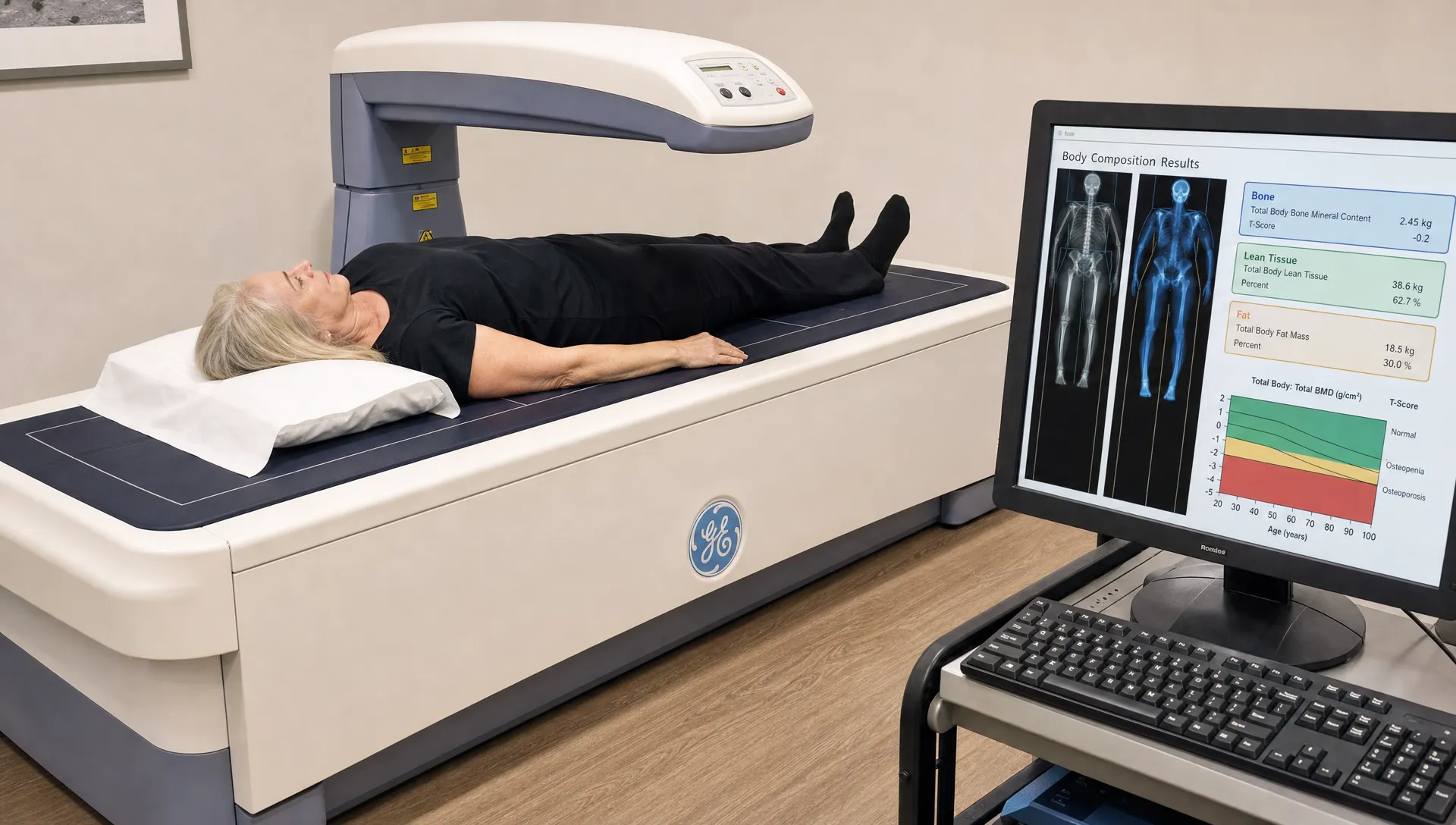
Fat mass is also estimated region by region. A full body result may include total fat mass, body fat percentage, trunk fat, arm and leg fat, android and gynoid regions and visceral fat estimates. This regional separation is one of the major reasons DEXA is more informative than a bathroom scale or body mass index.
Regional analysis is where the separation becomes practical
Whole-body body fat percentage is useful, but regional data often answers the better question. Did your legs gain lean mass after a strength block? Did your trunk fat decrease while scale weight stayed flat? Is one side carrying less lean mass than the other after an injury or sport-specific training pattern?
For training decisions, these details can reduce guesswork. If body weight is stable but fat mass is down and lean mass is up, the scale alone would miss that progress. If one limb is consistently lower in lean mass, that may guide unilateral strength work or a conversation with a coach or clinician. DEXA SF explains this applied side in more detail in its article on how a body scan for fat and muscle guides training.
What can affect the precision of tissue separation?
DEXA is highly useful, but it is still a measurement system. Results can be affected by hydration status, recent exercise, glycogen storage, food in the digestive tract, scan positioning and movement during the scan. These factors are especially relevant for lean mass because lean tissue includes water.
Standardization helps. The International Society for Clinical Densitometry publishes official positions that emphasize proper DXA acquisition, analysis and quality control for clinical use. For body composition tracking, the same general idea applies: consistent technique makes trend data more meaningful.
Practical ways to improve comparability include:
- Schedule repeat scans under similar conditions when possible.
- Avoid comparing a scan after hard training with one taken after several rest days.
- Remove metal items and follow the facility’s positioning instructions.
- Use the same scan provider and report format when tracking change over time.
Why repeat scans should be compared carefully
Over time, dual energy imaging is most valuable when you look for patterns rather than overreacting to one small change. A modest shift in lean mass after a salty meal, heavy workout or different hydration state may not reflect true muscle gain or loss. A larger trend across standardized scans is more meaningful.
This is also why expert interpretation matters. A DEXA report may show many numbers, but not every number deserves the same attention. The right interpretation connects the scan result to your goal, whether that goal is fat loss, strength performance, bone health or maintaining lean mass during a nutrition phase.
From separated tissues to decisions you can act on
In practice, dual energy imaging matters because it turns one scan into several distinct outputs. Instead of asking whether weight went up or down, you can ask what changed. That distinction is especially valuable during body recomposition, where fat mass and lean mass may move in opposite directions.
| Result on a DEXA report | What was separated | How it can guide decisions |
|---|---|---|
| Body fat percentage | Fat mass compared with total mass | Shows whether weight change is coming from fat, lean tissue or both |
| Lean mass by region | Non-fat, non-bone tissue in arms, legs and trunk | Helps track training response and possible asymmetries |
| Bone mineral density | Mineral signal separated from soft tissue | Supports bone health discussions and long-term monitoring |
| Visceral fat estimate | Deep abdominal fat estimated from regional data | Adds context beyond total body fat percentage |
| Android to gynoid pattern | Trunk and hip-leg fat distribution | Helps interpret where fat is stored, not just how much exists |
A knowledgeable review helps you avoid common mistakes, such as treating lean mass as pure muscle or focusing on body fat percentage while ignoring total fat mass. If you already have a report, this guide to DEXA scan interpretation can help you understand the major metrics before your next scan.
For many people, the biggest benefit is emotional clarity. Scale weight can be noisy. DEXA does not remove all uncertainty, but it gives you a better map of what is changing inside that weight.
Frequently Asked Questions
Is DEXA the same as a regular X-ray? No. A regular X-ray is designed mainly to show anatomy and structural differences, especially bone. DEXA uses two X-ray energy levels and calibrated analysis to estimate bone mineral, fat mass and lean soft tissue.
Does dual energy imaging measure muscle directly? Not directly. It estimates lean soft tissue, which includes muscle but also water, organs, connective tissue and other non-fat tissue. For fitness tracking, regional lean mass can still be a useful proxy for muscle-related change when scans are performed consistently.
Can DEXA separate visceral fat from other fat? DEXA can estimate visceral fat in the abdominal region using validated software models. It is not the same as directly imaging every fat depot with CT or MRI, but it adds useful context beyond total body fat percentage.
How often should I repeat a DEXA scan? The best interval depends on your goal. Someone in an active fat loss or training phase may scan more often than someone monitoring long-term bone and body composition trends. The key is to repeat scans under similar conditions so changes are easier to interpret.
Schedule a precise body composition scan in San Francisco
If you want more than a scale weight estimate, a professional DEXA body composition scan can show how fat, lean mass, bone density, visceral fat and regional asymmetries fit together. At DEXA SF, clients receive precise scan data along with expert interpretation, fitness guidance and nutrition advice so the numbers are easier to apply to real goals.