
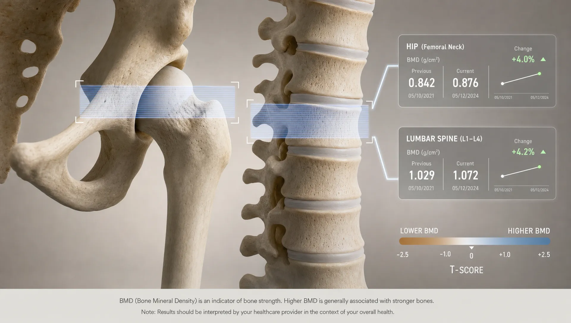
How DEXA Measures Bone Mineral Density at the Hip and Spine
Bone mineral density sounds like a single number, but a DEXA scan measures it at specific skeletal sites, most often the hip and lumbar spine. Those two areas matter because they are common fracture sites, they respond to age and hormone changes and they give clinicians standardized reference points for assessing bone health over time.
A DEXA scan for bone mineral density is not just a picture of the skeleton. It is a calibrated measurement that combines low-dose X-ray imaging, anatomical positioning, software-defined regions and comparison data. Understanding how the hip and spine are measured makes your results easier to read and helps explain why small technique differences can affect follow-up scans.
If you are new to the appointment itself, DEXA SF has a practical bone density scan guide that explains preparation and what to expect during the visit. This article focuses on the measurement method behind the numbers.
What DEXA is actually measuring
DEXA stands for dual-energy X-ray absorptiometry. You may also see it written as DXA. The scanner sends two low-dose X-ray beams through the body at different energy levels. Because bone and soft tissue absorb those beams differently, the system can estimate how much mineral content is present in the bone region being scanned.
The main output is bone mineral density, usually shown as grams per square centimeter, or g/cm². Technically, DEXA measures areal BMD, meaning it calculates mineral content over a two-dimensional projected area. It is not the same as volumetric density from a CT scan, but DEXA is widely used because it is fast, standardized, low dose and supported by large reference databases.
A DEXA report may include several related values:
- BMC: Bone mineral content, the estimated amount of mineral in the selected region.
- BMD: Bone mineral density, calculated from mineral content divided by projected bone area.
- T-score: Your BMD compared with a healthy young adult reference group.
- Z-score: Your BMD compared with what is expected for someone of similar age and sex.
The International Society for Clinical Densitometry provides official positions for how central DEXA should be performed and interpreted. For diagnosis in most adults, the key central sites are the lumbar spine and proximal femur, which includes the femoral neck and total hip.
Why the hip and spine are the main DEXA sites
The hip and spine are measured because they carry high clinical value. Hip fractures are strongly associated with loss of independence and medical complications in older adults. Vertebral fractures can cause pain, height loss and posture changes, yet they may occur without being recognized right away.
These sites also show different aspects of bone health. The lumbar spine contains more trabecular bone, a porous type of bone that can change relatively quickly with aging, menopause, some medications and treatment. The hip includes both cortical and trabecular bone and is central to fracture risk assessment.
Measuring both areas gives a more complete view than either one alone. Some people lose density first at the spine, others show greater loss at the hip. A scan can also reveal discordance, meaning one site looks normal while another falls into low bone mass or osteoporosis range. That difference is one reason a clinician should interpret your report in context rather than relying on a single number.
If you are deciding whether testing makes sense for you, this overview of when to get a bone DEXA scan covers common timing and risk factors.
How DEXA measures bone mineral density at the hip
For the hip scan, the technologist positions one hip, often the left unless surgery, hardware or another factor makes the other side more suitable. The leg is gently rotated inward so the femoral neck is displayed correctly. This detail matters because rotation changes how the hip anatomy projects on the scan and can alter the region the software measures.
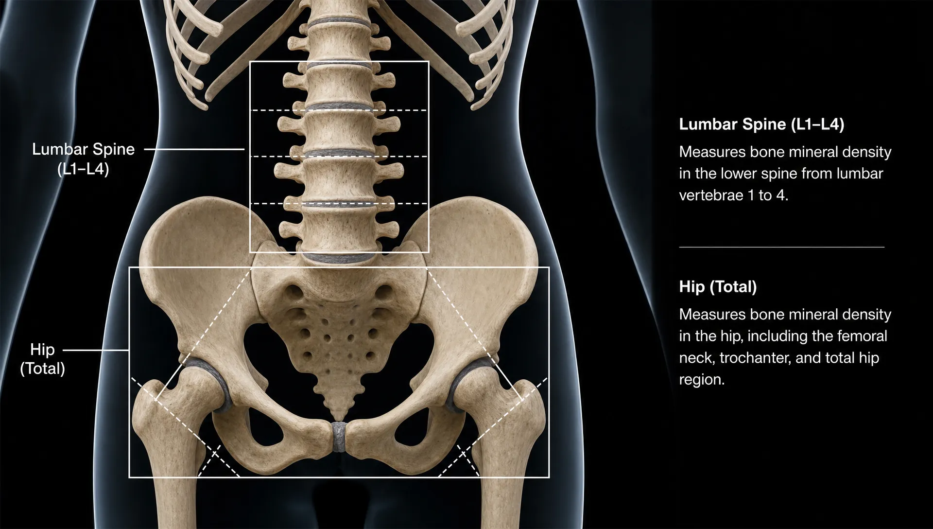
The scanner captures the proximal femur, which is the upper part of the thigh bone near the pelvis. The analysis software identifies anatomical landmarks, then places regions of interest over standardized areas. The two most important diagnostic regions are the femoral neck and the total hip.
| Hip region | What it measures | Why it matters |
|---|---|---|
| Femoral neck | Narrow area just below the ball of the hip joint | Common site used in fracture risk tools and osteoporosis classification |
| Total hip | Broader region including the femoral neck, trochanter and intertrochanteric area | Often more reproducible for monitoring change over time |
| Trochanter | Outer upper portion of the femur | May appear on reports, but is not usually the primary diagnostic site |
For diagnosis, official positions generally use the lower valid T-score from the femoral neck or total hip. Ward's area, a small region within the femoral neck, may appear on some older reports but is not used for diagnosing osteoporosis.
The hip is valuable because it is less affected than the spine by some age-related artifacts, such as degenerative changes. That does not mean it is immune to technical issues. Prior hip replacement, metal hardware, severe arthritis or poor positioning can make a hip result less reliable. In those cases, the other hip or another skeletal site may be needed.
How DEXA measures bone mineral density at the spine
The standard spine measurement is taken from the lumbar spine, usually vertebrae L1 through L4, with the person lying on their back. The technologist may elevate the legs on a support to reduce the natural curve of the lower back and improve positioning.
The scan is typically a posterior-anterior view, meaning the X-ray beam passes through the body to create a front-facing projection of the lumbar vertebrae. The software outlines the vertebral bodies, separates them into L1, L2, L3 and L4, then calculates BMD across the evaluable vertebrae.
The lumbar spine is sensitive to changes in trabecular bone. That makes it useful for detecting early bone loss and monitoring treatment response. It also means the spine can show improvement or decline before the hip in some people.
Spine interpretation requires careful review because the lower back often develops changes that can falsely raise BMD. Osteoarthritis, bone spurs, aortic calcification, scoliosis, vertebral compression fractures or surgical hardware can interfere with the result. When a vertebra is affected by a clear artifact or structural abnormality, it may be excluded from the calculation according to accepted interpretation rules. If too few vertebrae remain valid, the clinician relies more heavily on another site.
What T-scores and Z-scores mean on a hip and spine report
Your BMD value is the raw density measurement. The T-score and Z-score put that measurement into context.
A T-score compares your BMD with the average peak bone density of a healthy young adult reference population. It is commonly used for osteoporosis classification in postmenopausal women and men age 50 and older. A Z-score compares your BMD with an age-matched reference group and is often more relevant in younger adults, premenopausal women and children.
The World Health Organization diagnostic categories are commonly summarized this way for T-scores:
| Category | T-score range | General meaning |
|---|---|---|
| Normal bone density | -1.0 or higher | BMD is within the expected young adult reference range |
| Low bone mass, also called osteopenia | Between -1.0 and -2.5 | BMD is below normal but not in the osteoporosis range |
| Osteoporosis | -2.5 or lower | BMD meets the densitometric threshold for osteoporosis |
These categories are useful, but they do not replace medical judgment. The Bone Health and Osteoporosis Foundation Clinician's Guide emphasizes that fracture history, age, medications, fall risk and other clinical factors also matter when deciding what to do next.
Two people can have the same T-score and different fracture risk. For example, a person with a prior fragility fracture, long-term glucocorticoid use or frequent falls may need a different clinical plan than someone with the same BMD and no other major risk factors.
Why positioning and consistency matter
DEXA is precise, but it is not magic. The quality of a hip or spine result depends on the scanner, calibration, positioning and analysis. That is why trained technologists and consistent protocols are so important.
For the hip, the leg should be positioned so the femoral neck is clear and comparable with reference standards. Too much or too little rotation can change the apparent shape of the femur and affect the measured region.
For the spine, the vertebrae should be centered and straight enough for accurate analysis. If the spine is tilted, rotated or partly outside the field, the software may place boundaries incorrectly. A qualified reviewer can correct some analysis issues, but a poorly positioned scan may need to be repeated.
Consistency is especially important when tracking change. If you compare one scan with a future scan, the best comparison usually comes from the same facility, same machine type and same measurement sites. Clinicians also consider the least significant change, which is the minimum change needed to be confident that BMD has truly changed rather than shifted because of normal measurement variation.
This is why a small difference on a follow-up report does not always mean bone density has meaningfully improved or worsened. The interpretation depends on the size of the change, the site measured and the facility's precision standards.
Common reasons hip and spine results do not match
It is common for the hip and spine to show different categories. One site may be normal while another shows low bone mass, or one may meet the osteoporosis threshold while the other does not.
Several factors can explain this pattern:
- Different bone types: The spine has more trabecular bone, while the hip has a different mix of cortical and trabecular bone.
- Aging pattern: Bone loss may appear earlier or faster at one site depending on hormones, activity, medication history and genetics.
- Artifacts: Spine arthritis or calcification can make lumbar BMD appear higher than it really is.
- Prior injury or surgery: Hardware, fractures or joint replacement can make a site unsuitable for interpretation.
- Mechanical loading: Strength training, body weight and movement patterns can influence bone differently across regions.
When sites disagree, clinicians usually consider the lowest valid diagnostic site along with the person's overall risk profile. They may also look at prior scans to see whether the pattern is stable or changing.
How DEXA bone density fits with body composition
A central bone density scan focuses on the hip and spine, but DEXA technology can also measure body composition. Depending on the scan protocol, DEXA can assess fat mass, lean mass, visceral fat and left-right asymmetries. Those measurements do not diagnose osteoporosis, but they can help you understand the physical context around bone health.
Muscle and bone are connected. Resistance training can help maintain muscle mass and place beneficial load on bone. Nutrition also matters, particularly adequate protein, calcium, vitamin D and total energy intake. If you are trying to interpret bone results alongside body composition, DEXA SF's article on body and bone health explains what one scan can show beyond weight alone.
A DEXA report should not be treated as a standalone prescription. It is a measurement tool. The next step may involve reviewing results with a clinician, especially if your T-score is low, your Z-score is unexpectedly low for your age or you have a history of fractures. Fitness and nutrition guidance can support healthier habits, but medical diagnosis and treatment decisions belong with a qualified healthcare professional.
What to look for when reviewing your report
When you receive a DEXA report, start with the measured sites. Confirm whether the report includes lumbar spine, femoral neck and total hip values. Then look at which site has the lowest valid T-score, since that often drives classification.
Next, check whether any vertebrae were excluded from the lumbar spine analysis. An exclusion is not necessarily a problem. It may mean the interpreter removed an artifact to make the result more accurate. If several vertebrae are excluded, ask which site should be used for follow-up comparison.
For repeat scans, compare the same region over time. Total hip should be compared with total hip, femoral neck with femoral neck and lumbar spine with lumbar spine. Mixing sites can make trends look more dramatic or more reassuring than they really are.
Finally, read the impression or interpretation section, not just the numbers. The impression should place the BMD findings into categories and may mention whether the change from a prior scan is significant.
Frequently Asked Questions
Is a DEXA scan for bone mineral density the same as a full body composition scan? It uses the same core technology, but the protocol and report can differ. A diagnostic bone density scan focuses on central skeletal sites such as the hip and spine. A body composition DEXA scan measures fat mass, lean mass and other composition metrics, and may include bone data that is useful for wellness tracking.
Why does DEXA measure the hip and spine instead of every bone? The hip and spine are standardized central sites with strong clinical relevance for fracture risk and osteoporosis classification. Measuring every bone would add time and complexity without improving routine diagnosis for most people.
Can arthritis make my spine bone density look higher? Yes. Degenerative changes, bone spurs and calcification near the lumbar spine can falsely increase the measured spine BMD. This is one reason the hip result and the scan images themselves are reviewed, not just the spine number.
Which hip number matters most, femoral neck or total hip? Both matter. For diagnosis, clinicians generally use the lower valid T-score from the femoral neck or total hip. For monitoring over time, the total hip is often useful because it tends to be more reproducible.
How often should hip and spine DEXA scans be repeated? Timing depends on your age, baseline result, risk factors and whether you are being monitored during treatment. Many people repeat testing after one to two years when there is a clinical reason, but your clinician should set the interval.
Measure bone density with more context
A DEXA scan can turn bone health from a vague concern into measurable data. At DEXA SF, scans can help assess bone density alongside body fat, lean mass, visceral fat, asymmetries and metabolic insights, with expert interpretation and practical fitness and nutrition guidance.
If you want a clearer baseline for your bone and body composition, you can schedule a DEXA scan with DEXA SF. Bring your results to your healthcare provider if you need medical diagnosis, treatment decisions or osteoporosis management.