
Can a Bone Density X-Ray Detect Early Bone Loss?
A bone density X-ray can detect early bone loss when the test is a DEXA scan, also called DXA. That distinction matters. A standard X-ray can sometimes show severe bone thinning, but it is not sensitive enough to reliably catch early bone loss. DEXA is different because it measures bone mineral density directly and compares your results with reference ranges used to identify osteopenia and osteoporosis.
For people who feel healthy, train regularly or have no obvious symptoms, this can be surprising. Bone loss usually does not cause pain until a fracture happens. A DEXA scan helps find risk earlier, when there may still be time to adjust nutrition, resistance training, medications or other clinical strategies with your healthcare provider.
What people usually mean by a bone density X-ray
The phrase “bone density X-ray” is often used casually, but it can refer to two very different things.
A conventional X-ray creates a picture of bones, joints or an injured area. It is useful for finding fractures, arthritis changes, alignment problems and certain bone abnormalities. It can show bones that look unusually thin, but only after density loss is more advanced.
A DEXA scan uses low dose X-ray beams to estimate bone mineral density. Instead of simply producing an image, it calculates how much mineral content is present in specific bone regions, most often the hip and spine. That number is then compared with reference populations to estimate whether your bone density is normal, low or in the osteoporosis range.
If you are new to the terminology, DEXA SF has a helpful guide that explains what a DEXA X-ray is for first-time patients, including how it differs from a regular X-ray.
Can it detect early bone loss?
Yes, a DEXA bone density X-ray can detect early bone loss before it would typically be visible on a regular X-ray. This early stage is often called low bone mass or osteopenia.
DEXA does this by measuring bone mineral density, usually reported in grams per square centimeter, then translating that result into scores your clinician can interpret. The most familiar is the T-score.
| T-score range | What it generally means |
|---|---|
| -1.0 or higher | Normal bone density |
| Between -1.0 and -2.5 | Low bone mass, often called osteopenia |
| -2.5 or lower | Osteoporosis range |
These categories are commonly used in adults age 50 and older. For younger adults, clinicians often pay closer attention to Z-scores, which compare bone density with what is expected for someone of the same age and sex.
This is where DEXA becomes valuable for early detection. Someone can feel completely well, have no fracture history and still have a T-score that shows bone loss has already started. A regular X-ray would usually not provide that level of detail.
Why a regular X-ray is not enough for early bone loss
A regular X-ray depends on visible contrast. Mild bone loss often does not change the image enough for a radiologist to identify it confidently. By the time bones look clearly “thin” on a standard X-ray, the person may already have substantial bone mineral loss.
The American College of Radiology and Radiological Society of North America patient resource RadiologyInfo.org describes DEXA as the most commonly used test to diagnose osteoporosis and assess fracture risk. That is because DEXA is quantitative. It gives a measured result, not just a visual impression.
A standard X-ray still has an important role. If you have pain after a fall, sudden back pain or a suspected fracture, your clinician may order a regular X-ray first. But if the question is “Am I losing bone density early?” a DEXA scan is the more appropriate tool.
What a DEXA scan measures beyond the image
A DEXA scan is not just a picture of your bones. It measures bone mineral density at clinically meaningful sites and produces scores that help classify risk. Depending on the type of scan and the provider, DEXA can also measure body composition, including fat mass, lean mass, visceral fat and left vs. right muscle asymmetry.
For bone health, the key outputs often include:
- Bone mineral density: The measured mineral content in scanned bone regions.
- T-score: A comparison with the average bone density of a healthy young adult reference population.
- Z-score: A comparison with people of similar age, sex and body size.
- Regional data: Results from areas such as the hip, femoral neck or lumbar spine.
If you want a broader view of what the scan can report, this guide to what a BMD scan measures and when to get one explains the main results in more detail.
Why early bone loss is easy to miss
Bone is living tissue. It is constantly being broken down and rebuilt. In younger adulthood, bone formation and bone breakdown are usually balanced. With aging, menopause, certain medications, low energy intake, low vitamin D, endocrine conditions or reduced strength training, breakdown can outpace rebuilding.
The problem is that early bone loss usually has no obvious symptoms. You cannot feel your T-score dropping. You may not notice any change in strength, posture or daily function. Many people first learn they have low bone density after a fracture from a fall that seemed too minor to cause one.
The National Institute on Aging notes that osteoporosis is sometimes called a “silent disease” because people may not know they have it until a bone breaks. DEXA helps shift the timeline earlier by identifying low bone density before a fracture forces the issue.
What happens during a DEXA bone density scan
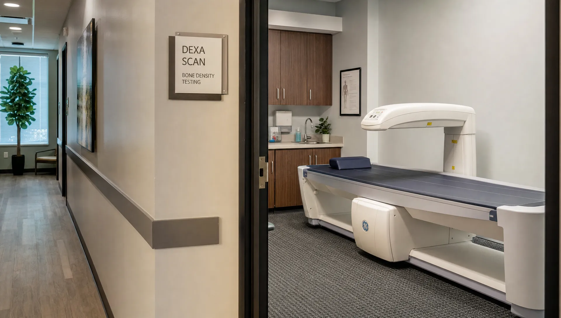
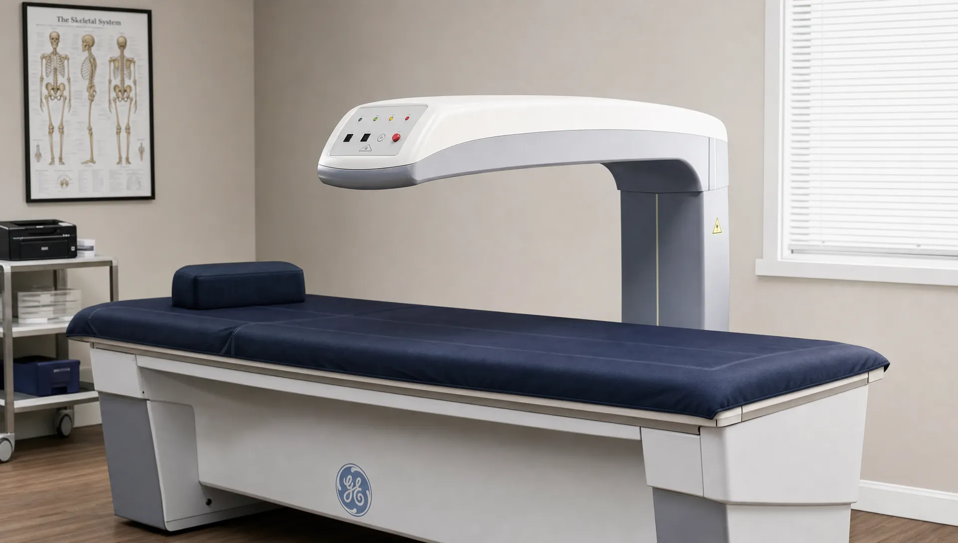
A DEXA scan is typically quick and noninvasive. You lie still on a scanning table while the machine passes over the body. For a clinical bone density scan, the most common measurement sites are the hip and spine because they are strongly related to osteoporotic fracture risk.
You do not feel the X-rays during the scan. The radiation exposure from DEXA is low, much lower than many conventional imaging tests. You may be asked to avoid calcium supplements for a short period before the scan or wear clothing without metal, but preparation is usually simple. Always follow the instructions from the imaging provider or your clinician.
The result should be interpreted in context. A T-score is useful, but it is not the whole story. Age, fracture history, family history, medications, smoking, alcohol intake, body weight, balance, fall risk and medical conditions can all affect overall fracture risk.
Who should consider screening for early bone loss?
Screening recommendations vary by age, sex and risk profile. The U.S. Preventive Services Task Force recommends screening women age 65 and older for osteoporosis, and screening postmenopausal women younger than 65 who are at increased risk. For men, the USPSTF states that evidence is insufficient to make a broad screening recommendation, so individual risk discussion with a clinician is especially important.
A clinician may consider earlier bone density testing if you have risk factors such as:
- A prior fracture from a low impact fall
- Long-term glucocorticoid use
- Early menopause or low estrogen exposure
- Low body weight or a history of undernutrition
- Smoking or heavy alcohol use
- Rheumatoid arthritis, hyperthyroidism or other conditions linked with bone loss
- A parent with hip fracture
- Significant height loss or possible vertebral compression fracture
Athletes and highly active people are not automatically protected. Resistance training supports bone health, but low energy availability, restrictive dieting, missed menstrual cycles, low vitamin D or endurance training without enough loading can raise risk in some individuals.
What early detection can change
The value of early detection is not only knowing a number. It is using that number to make better decisions.
If a DEXA scan shows early bone loss, your clinician may evaluate vitamin D status, calcium intake, hormone status, medications or underlying medical conditions. A fitness professional or qualified health expert may also help you design a safer strength training plan that applies progressive loading without unnecessary injury risk.
Nutrition can matter too. Bone needs adequate protein, calcium, vitamin D and total energy intake. For some people, the issue is not one missing nutrient but a long-term mismatch between training demands and recovery. For others, medication review or medical treatment may be appropriate.
Follow-up scans can show whether your plan is working. Bone changes slowly, so repeat testing is usually spaced out rather than done every few weeks. The right interval depends on baseline results, risk factors and clinical decisions.
DEXA versus other bone tests
A DEXA scan is sometimes confused with a “bone scan,” but they are not the same. A nuclear medicine bone scan looks at bone metabolism and is used for problems such as certain fractures, infection, cancer spread or unexplained bone pain. It is not designed to measure bone mineral density for osteoporosis screening.
Quantitative CT can measure bone density too, but it is less commonly used for routine screening and usually involves more radiation than DEXA. Ultrasound heel tests may estimate risk in some settings, but they do not replace central DEXA measurements at the hip and spine when diagnostic classification is needed.
For early bone loss, DEXA remains the standard test because it is validated, widely used, relatively quick and specifically designed to quantify bone density.
How to read results without overreacting
A low T-score deserves attention, but it should not be interpreted in isolation or used to self-diagnose. Bone density is one part of bone strength. Bone quality, fall risk, muscle mass, balance, vision, medications and overall health also influence fracture risk.
For example, two people can have the same T-score but different risk profiles. One may be 52, active, with no fracture history and no major risk factors. Another may be 78, taking medications that affect balance and have already had a wrist fracture. The same scan result can lead to different next steps.
This is why expert interpretation matters. A good scan experience should help you understand what the numbers mean, what they do not mean and what actions may be worth discussing with your healthcare provider.
Can improving muscle help protect bone?
Muscle and bone health are closely related. Muscle applies force to bone during movement, especially during resistance training and weight-bearing activity. Stronger muscles can also improve balance and reduce fall risk, which is a major part of fracture prevention.
DEXA body composition data can add useful context here. If someone has low lean mass, significant left-right asymmetry or high visceral fat, those findings may influence fitness and nutrition goals alongside bone density results. At DEXA SF, scans can provide data on body fat, lean mass, bone density, visceral fat and asymmetries, with expert interpretation to help connect the numbers to practical next steps.
For a combined look at these relationships, the DEXA SF article on body and bone health explains how one scan can show more than a single bone density number.
When a bone density X-ray may not be the right first step
A DEXA scan is useful for detecting low bone density, but it is not the answer to every bone-related concern. If you have acute pain after trauma, sudden swelling, unexplained severe back pain, fever, cancer history or neurological symptoms, contact a medical professional promptly. You may need a different evaluation before or instead of DEXA.
DEXA also does not explain every cause of bone pain. It can show low bone density, but it does not diagnose all fractures, infections, tumors or inflammatory conditions. The right test depends on the clinical question.
Frequently Asked Questions
Can a bone density X-ray detect osteopenia? Yes. A DEXA bone density X-ray can detect low bone mass, commonly called osteopenia, by measuring bone mineral density and calculating a T-score.
Can a regular X-ray detect early osteoporosis? Usually not reliably. A regular X-ray may suggest bone thinning when loss is more advanced, but DEXA is the preferred test for measuring bone density and detecting earlier loss.
Is a DEXA scan painful? No. A DEXA scan is noninvasive. You lie still on a table while the scanner passes over the body, and you do not feel the X-rays.
How often should bone density be checked? The right interval depends on your age, baseline results, risk factors and whether treatment is being monitored. Your clinician can recommend a schedule based on your situation.
Does a normal DEXA result mean I have no fracture risk? Not necessarily. A normal result is reassuring, but fracture risk also depends on falls, medications, muscle strength, balance, vision and other health factors.
Get clearer answers about bone and body composition
A bone density X-ray can detect early bone loss when it is a DEXA scan, not a conventional X-ray. That difference can help you catch low bone density before symptoms or fractures appear.
If you are in San Francisco and want precise data on bone density, body fat, lean mass, visceral fat and asymmetries, DEXA SF provides professional DEXA scans with expert interpretation, plus fitness and nutrition guidance based on your results. Schedule a scan to understand where you are now and what to discuss next with your healthcare provider.