
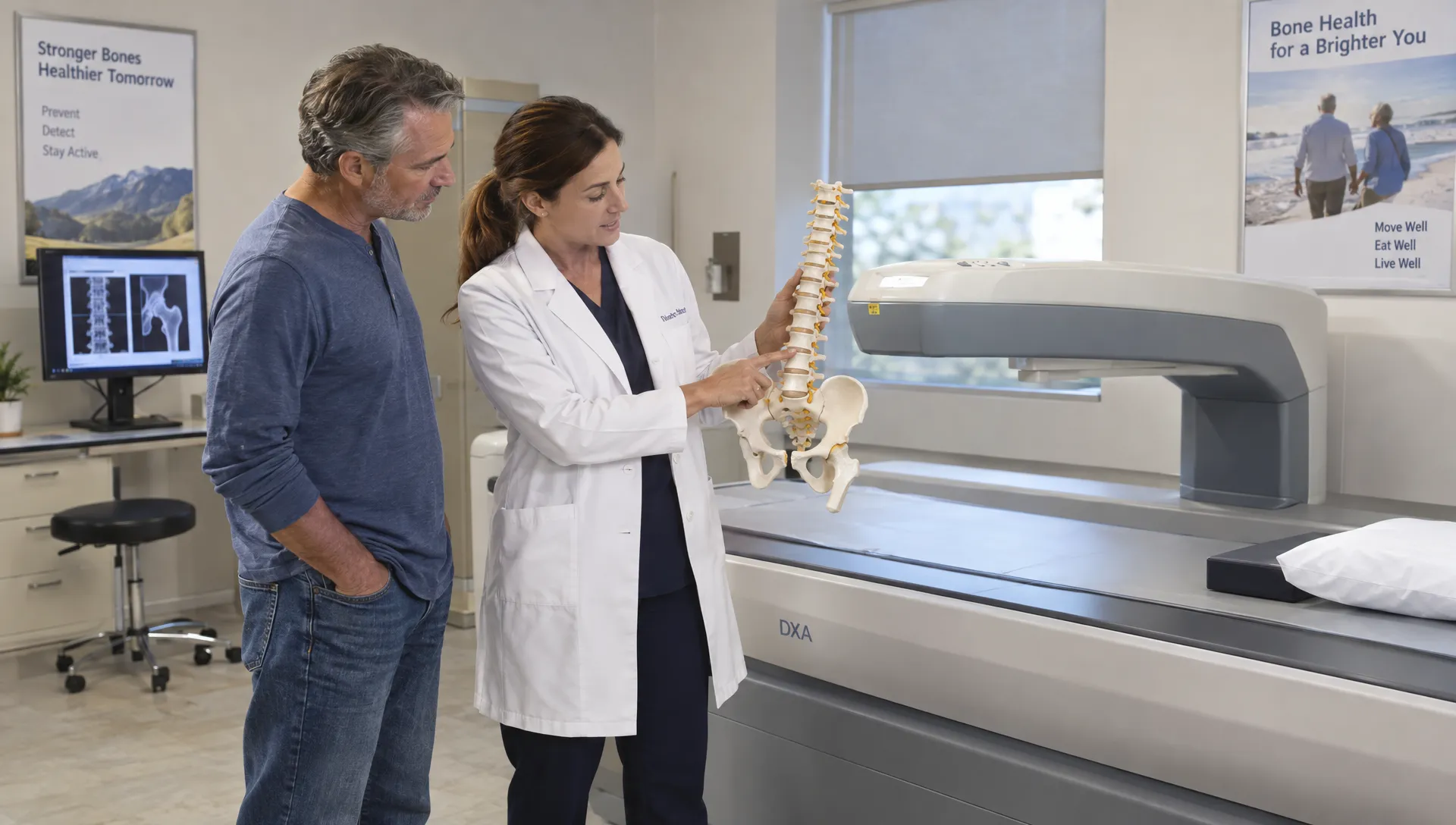
When Arthritis Can Affect a Bone Densitometry Scan
Arthritis can affect a bone densitometry scan in two different ways: degenerative changes may make bone density appear higher than it really is, while inflammatory arthritis can cause genuine bone loss. Both can happen in the same person. That means a reassuring spine result does not always tell the whole story.
Having arthritis does not make bone density testing useless. It makes the choice of measurement sites, review of the scan images and interpretation of the results especially important. Here is when arthritis matters and what to discuss with your healthcare team.
Two ways arthritis can change the result
A DXA scan, also called DEXA, uses low-dose X-rays to measure bone mineral density. A diagnostic osteoporosis examination typically evaluates the lumbar spine and hip. It measures mineral within a projected area, rather than directly testing bone strength or examining joint cartilage.
Arthritis can complicate that measurement through measurement interference, such as bone spurs overlapping the area being analyzed. Separately, some forms of arthritis increase the risk of actual bone loss through inflammation, reduced activity or medication effects.
These are different problems. One calls for careful image interpretation; the other may also require evaluation and treatment of osteoporosis risk.
When osteoarthritis affects a bone densitometry scan
Osteoarthritis is the form most likely to make lumbar spine measurements look artificially reassuring. Wear-related changes can produce osteophytes, commonly called bone spurs, and areas of denser bone called sclerosis. These changes can contribute mineral to the area the scanner measures without indicating stronger vertebral bone.
The effect is particularly relevant to the standard front-to-back lumbar spine measurement. Degeneration in the spine, including the facet joints, can influence the reading. A person can therefore have substantial spinal arthritis and a relatively high spine bone density result while having low density at the hip.
A higher spine reading caused by degeneration is not the same as protection against fracture. Osteoarthritis and osteoporosis can coexist, and the presence of one does not exclude the other.
The amount of interference varies. Mild arthritis may have little effect, while more extensive changes can make particular vertebrae unsuitable for interpretation.
Findings that deserve a closer look
Arthritis is not the only explanation for an unexpectedly high or uneven spine result. Other findings can also complicate interpretation.
| Finding | How it can affect interpretation | What the interpreting clinician may review |
|---|---|---|
| Osteophytes and spinal sclerosis | May raise measured lumbar density | Individual vertebrae and the scan image |
| A compressed vertebra | May produce an unexpectedly high density measurement because its projected area changes | Fracture history and additional imaging when indicated |
| Spinal hardware | Can interfere with the measured region | Whether the affected region should be excluded |
| Marked aortic calcification | Can sometimes contribute to an elevated front-to-back spine reading | The location of calcification relative to the measurement |
These findings do not automatically invalidate a bone densitometry scan. They are reasons to examine the underlying images rather than relying only on the summary score.
Inflammatory arthritis can cause genuine bone loss
Rheumatoid arthritis presents a different concern. Persistent inflammation can increase bone breakdown, both near affected joints and throughout the skeleton. Pain, fatigue and reduced mobility may also limit weight-bearing activity and contribute to bone loss.
Glucocorticoids such as prednisone add another risk. They can weaken bone, and fracture risk may be higher than a density result alone suggests. The National Institute of Arthritis and Musculoskeletal and Skin Diseases’ osteoporosis overview explains the role of medical conditions and medications in bone loss.
Not every arthritis medication has this effect. Do not assume that all disease-modifying treatments weaken bones, and do not stop prescribed medication before a scan unless your clinician instructs you to do so.
For someone with inflammatory arthritis, a bone densitometry scan should be interpreted alongside the diagnosis, medication exposure, prior fractures and other risk factors. A result outside the osteoporosis range does not necessarily mean fracture risk is low.
How clinicians obtain useful results despite arthritis
The goal is not to discard every measurement affected by age-related change. It is to identify which measurements remain reliable and use established interpretation standards.
Review individual lumbar vertebrae
A diagnostic lumbar spine examination generally analyzes the L1 through L4 vertebrae. The interpreting clinician reviews them for structural abnormalities, artifacts and unusual differences between neighboring levels.
The International Society for Clinical Densitometry’s Adult Official Positions explain when affected vertebrae should be excluded. If too few evaluable vertebrae remain, diagnosis should rely on another valid skeletal site rather than a single remaining vertebra.
Patients should not try to remove a vertebra from the calculation themselves. An unusual value is a reason for professional review, not proof that the number is wrong.
Use an interpretable hip or forearm measurement
When spinal degeneration limits interpretation, hip measurements often provide useful information. Arthritis can affect the hip too, so the image, positioning and presence of any joint replacement still matter.
If one hip has been replaced, the other hip may remain suitable. When the hip and spine cannot be measured or interpreted adequately, a forearm measurement may be appropriate. The standard diagnostic forearm region is the one-third radius, usually on the nondominant side.
For a bone densitometry scan affected by arthritis, choosing an alternative site is a clinical decision, not simply a matter of selecting whichever number looks lowest. Different skeletal regions have defined roles in osteoporosis assessment.
Check whether follow-up changes are believable
An increase in lumbar density over time does not always mean treatment has strengthened the bone. Progressing degeneration can raise the reading, while changes in positioning or the vertebrae included in the analysis can also affect comparisons.
Whenever possible, follow-up testing should use the same facility and equipment, or appropriately cross-calibrated systems. The interpreting team should assess whether a difference exceeds the facility’s least significant change, the threshold used to distinguish a measurable change from testing variability.
A useful question is: “Does this change represent bone improvement, or could arthritis or a technical difference explain it?” That is especially relevant when spine density rises while hip density stays stable or falls.
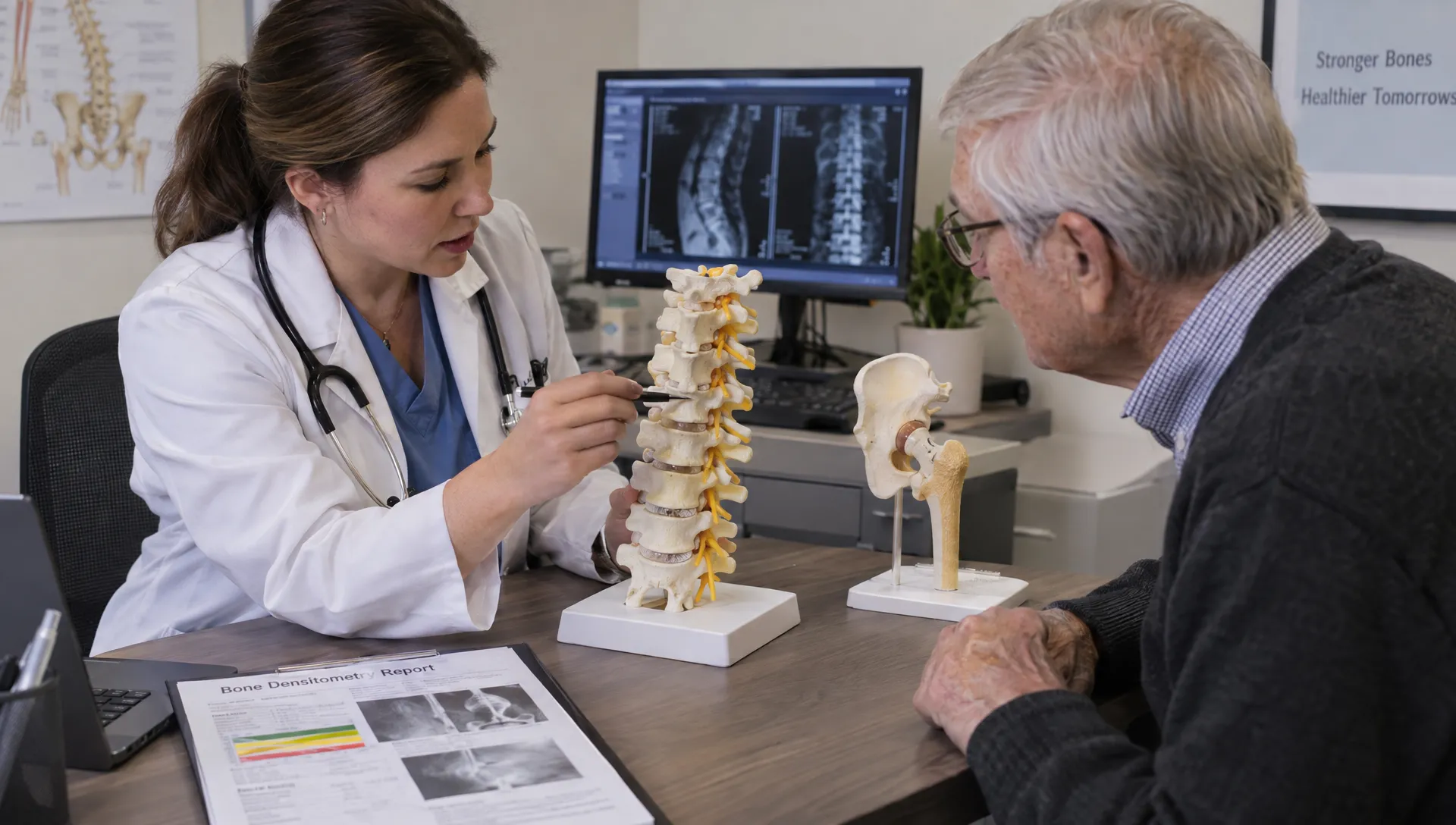
What to tell the imaging team before your visit
You usually do not need to cancel testing because you have arthritis. Instead, give the team enough information to plan the examination and interpret it appropriately.
Before your bone densitometry scan, mention the following:
- Your arthritis diagnosis and whether your spine or hips are affected.
- Any hip replacements, spinal fusion, metal hardware or previous spine surgery.
- Known vertebral compression fractures, unexplained height loss or recent back pain.
- Current or previous prednisone use, including the dose and duration if you know them.
- Pain or stiffness that makes lying flat or rotating a leg difficult.
- Previous bone density reports and where those examinations were performed.
Do not force a painful position. Tell the technologist about limitations before positioning begins so they can assess whether adjustments or another measurement site are needed.
Medication history matters beyond arthritis treatment. Our guide to medications that may affect a bone mineral density test explains why the team needs to distinguish long-term effects on bone from factors that may interfere with the examination. Follow the facility’s preparation instructions rather than changing treatment on your own.
How to interpret a report that mentions degeneration
Look for more than the word “normal.” A useful report identifies the sites measured, notes important limitations and explains whether any vertebrae were excluded.
For postmenopausal women and men age 50 or older, a T-score of −2.5 or below at an appropriate diagnostic site can establish densitometric osteoporosis. Younger adults are generally evaluated using Z-scores and clinical context, rather than applying the same T-score classification automatically.
When arthritis affects a bone densitometry scan, the main question is whether the reported score comes from a valid, interpretable region. Spine and hip results should not simply be averaged to make an abnormal result disappear.
Clinicians may also consider a fracture-risk assessment such as FRAX, which includes rheumatoid arthritis and glucocorticoid exposure among its risk factors. FRAX has limitations and does not replace clinical judgment, particularly for people with recent fractures or substantial steroid exposure.
A low-trauma fracture can remain important even when the density score is not in the osteoporosis range. Sudden back pain after a minor movement, new height loss or a suspected fracture deserves medical assessment rather than reassurance from a density number alone.
Confirm which type of DEXA examination you need
A whole-body composition scan and a dedicated osteoporosis examination use related technology, but they are not automatically interchangeable. A whole-body bone measurement should not be assumed to replace diagnostic lumbar spine and hip measurements.
If your concern is arthritis-related fracture risk, ask whether the planned examination includes the appropriate diagnostic sites and medical interpretation. Our overview of when to get a bone DEXA scan and what it shows provides additional context for that conversation.
Frequently asked questions
Can I have osteoporosis even if arthritis makes my spine score look normal? Yes. Degenerative changes can raise the measured spine value without eliminating osteoporosis elsewhere. Hip measurements, other appropriate sites and your fracture history may provide a more reliable assessment.
Can a bone densitometry scan diagnose arthritis? No. Its primary purpose is measuring bone mineral density, not diagnosing the cause of joint pain or assessing cartilage damage. Arthritis evaluation may involve a physical examination, laboratory testing and other imaging, depending on the symptoms.
Does rheumatoid arthritis always cause an abnormal result? No. Bone density varies with age, disease activity, treatment, mobility and other factors. Rheumatoid arthritis increases concern about bone health, but an individual’s result cannot be predicted from the diagnosis alone.
Should I postpone testing during an arthritis flare? Not automatically. Contact the imaging team if pain would prevent safe, reproducible positioning. They can advise whether to proceed, adjust the examination or coordinate with your referring clinician.
Choose the right scan for your bone health question
Arthritis is a reason to interpret bone density carefully, not a reason to avoid testing. Before booking, clarify whether you need body composition information, a diagnostic osteoporosis evaluation or both.
DEXA SF provides professional body composition scanning in San Francisco, with expert interpretation and personalized fitness and nutrition guidance. If your main concern is osteoporosis or an arthritis-related distortion in a previous result, discuss the required diagnostic examination with your healthcare clinician and confirm the scan type before scheduling.