

5 Body Scan Analysis Mistakes That Mislead Your Progress
Your body scan analysis shows a higher body fat percentage or less lean mass than last time. Before you cut calories or overhaul your workouts, check whether those numbers actually represent a change in your body. Different preparation, measurement variability and misread percentages can all make progress look worse than it is.
DEXA provides useful body composition measurements, but a report still needs interpretation. These five mistakes explain how an accurate scan can lead to an inaccurate conclusion, and what to check before acting on it.
1. Comparing scans taken under different conditions
A scan after breakfast and a hard workout is not directly comparable to one taken before breakfast on a rest day. Food, fluids, recent exercise and hydration can influence body composition results, especially measurements involving lean tissue.
The equipment and analysis process matter, too. Different scanners, software versions or positioning procedures can introduce differences that have nothing to do with your training program.
The misleading conclusion: “My new program caused me to lose lean mass.”
The better approach: First establish whether the two measurements were collected under sufficiently similar conditions. Consistent preparation makes body scan analysis more useful than trying to explain every difference after the fact.
Ask the scanning provider about its preparation instructions, then follow them consistently. Keep your usual hydration habits rather than deliberately restricting water or drinking extra to influence the result. Record anything unusual, such as a recent illness, travel or a demanding training session.
The International Society for Clinical Densitometry’s adult official positions identify preparation and technical factors that affect body composition measurement and recommend facility-specific precision assessment.
When possible, return to the same facility and scanner. If you switch locations, ask whether the results can be compared meaningfully. A different machine does not make the new scan useless, but it may be better treated as a new baseline rather than proof of a sudden change.
2. Treating every lean-mass change as muscle gained or lost
DEXA separates the body into fat mass, lean soft tissue and bone mineral content. Lean soft tissue includes skeletal muscle, but it also includes water, organs and other nonfat tissues. It is not a direct measurement of muscle fibers.
That distinction matters during dieting, changes in carbohydrate intake or a return to training. Glycogen is stored with water, so changes in glycogen stores can influence lean-tissue measurements without an equivalent change in muscle tissue.
The misleading conclusion: “My scan shows less lean mass, so I definitely lost that amount of muscle.”
The better approach: Interpret the lean-mass result alongside preparation, training performance and the longer-term trend. In body scan analysis, “lean mass” and “muscle mass” should not be treated as interchangeable labels.
Suppose your scan follows several days of lower carbohydrate intake and your lean mass is lower. That finding deserves attention, but it does not establish how much muscle you lost. If strength is stable and preparation differed from your previous appointment, the interpretation is less straightforward than the headline number suggests.
Conversely, an increase in lean mass after resuming training does not prove that the entire increase is new muscle. Look for agreement between repeat measurements, performance and other relevant observations.
Our explanation of what a body scan measures during weight loss explores this distinction further. Use the scan to identify a pattern worth investigating, not to assign an exact amount of muscle gain or loss from one comparison.
3. Judging progress by body fat percentage alone
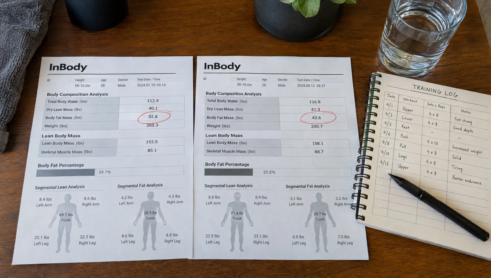
Body fat percentage is a ratio. It can change because fat mass changes, because other body components change or because both happen together.
This makes it a useful summary, but a poor standalone verdict. If you focus only on the percentage, you can miss the difference between gaining fat and having less nonfat mass.
Consider this simplified example, using fat mass divided by total body weight:
| Measurement | First assessment | Later assessment |
|---|---|---|
| Body weight | 80 kg | 78 kg |
| Fat mass | 24 kg | 24 kg |
| Calculated body fat percentage | 30% | Approximately 30.8% |
Fat mass did not increase. The percentage rose because total weight decreased. These illustrative figures explain the arithmetic, not a clinical target or the exact calculation used by every scan report.
The misleading conclusion: “My body fat percentage went up, so I gained fat.”
The better approach: Review absolute fat mass and lean mass before interpreting the percentage. Useful body scan analysis asks which component changed, rather than treating a ratio as the whole story.
The reverse problem is also possible. A lower percentage does not necessarily mean substantial fat loss if lean mass increased while fat mass stayed similar. That might still support your goal, but it is a different outcome.
Match the interpretation to your objective. During fat loss, examine whether fat mass decreased and lean tissue was reasonably maintained. During a muscle-building phase, look at lean-tissue trends alongside strength and the amount of fat gained. A single percentage cannot describe all of those outcomes.
4. Treating tiny differences in body scan analysis as definite progress
A report may display measurements to a decimal place, but that does not mean every decimal-place difference is meaningful. Measurement resolution and measurement precision are different things.
Precision describes how closely repeated measurements agree under comparable conditions. Even with careful preparation and a reliable scanner, some variation remains. Small changes in positioning or how regional boundaries are placed can also affect comparisons.
The misleading conclusion: “The number moved slightly, so my plan is definitely working,” or “One region decreased, so something is wrong.”
The better approach: Ask the facility how it evaluates measurement precision and whether the observed difference is large enough to distinguish from expected variability.
One useful concept is least significant change, a threshold derived from precision testing that helps determine whether a measured difference exceeds expected measurement variability. There is no single universal threshold that should be applied to every facility, metric and patient.
Reliable body scan analysis separates a detectable trend from a small fluctuation before recommending a major change. Exceeding a precision threshold supports confidence that a measurement changed, but it does not explain why the change occurred.
The interval between scans matters as well. Repeating a scan too soon may produce differences too small to interpret confidently. Ask your provider to recommend a follow-up interval based on your goal and the amount of change reasonably expected.
For regional results, avoid treating a small left-right lean-tissue difference as a diagnosis. Symptoms, strength testing and a clinical assessment may be more relevant when deciding whether an apparent imbalance needs attention.
5. Turning scan estimates into diagnoses or exact prescriptions
A detailed report can look more definitive than the measurements justify. Visceral fat estimates, bone measurements and metabolic figures each have a specific purpose, but they do not answer every health or nutrition question.
DEXA can estimate visceral adipose tissue using scanner software. That information may support a health discussion, but it does not independently diagnose diabetes or cardiovascular disease. Blood pressure, laboratory results, medical history and other clinical findings remain relevant.
Likewise, a whole-body composition scan is not automatically equivalent to a dedicated osteoporosis evaluation. Clinical bone density assessment commonly uses specific sites, particularly the hip and lumbar spine, with interpretation appropriate to the patient.
The misleading conclusion: “This report tells me exactly how many calories to eat and whether I have a medical condition.”
The better approach: Identify what was measured, what was estimated and what requires additional assessment. Good body scan analysis keeps those categories separate.
If a report includes resting metabolic rate calculated from body composition, that is an estimate, not a direct measurement of your energy expenditure. Resting needs also differ from total daily needs, which include activity and other energy costs. Indirect calorimetry is a separate method for measuring resting energy expenditure.
Use any calorie estimate as a starting point rather than an exact prescription. Your intake, activity, weight trend, recovery and goals help determine whether it fits your situation.
If a finding raises a health concern, ask which clinician or follow-up test is appropriate. Qualified interpretation should clarify the report’s limits as well as its useful findings.
A quick check before changing your plan
Before reacting to a scan comparison, establish three things: whether the measurements are comparable, whether the difference is meaningful and whether it supports a specific action.
| Check | What to verify |
|---|---|
| Scan conditions | Similar preparation, equipment and positioning |
| Metric definition | Fat mass, lean soft tissue, percentage or an estimate |
| Size of the change | Whether it exceeds expected measurement variability |
| Supporting evidence | Weight trend, training performance, symptoms and relevant clinical data |
| Next step | Continue monitoring, make a measured adjustment or seek clinical follow-up |
This check keeps body scan analysis focused on decisions rather than isolated numbers. A scan does not need to show improvement in every category to be useful, and an unexpected result does not automatically mean your plan failed.
If you need help organizing that discussion, our guide to reviewing your DEXA scan analysis with confidence explains how to connect report findings to your goals. Bring your previous report and relevant preparation notes so the comparison has context.
Frequently asked questions
Can dehydration affect a DEXA body composition scan? Yes. Changes in hydration can influence lean-tissue measurements and complicate comparisons. Follow your provider’s preparation instructions and maintain normal hydration rather than trying to manipulate the result.
How often should I repeat a scan to track progress? There is no universal interval. Choose one with your provider based on your goal, expected rate of change and measurement precision. Scanning too frequently can make small fluctuations look more important than they are.
Should I change my diet after one disappointing result? Not automatically. Body scan analysis should first establish whether the change is credible and relevant to your goal. Check preparation, absolute fat and lean mass, measurement variability and supporting trends before making a substantial adjustment.
Get a scan with interpretation, not just numbers
DEXA SF provides body composition scans in San Francisco with expert interpretation and personalized fitness and nutrition guidance. Bring your previous results and questions about any unexpected changes.
Schedule a DEXA scan with DEXA SF to establish a baseline or review your progress with the context needed to make a useful next decision.